 GCEO Group®
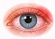 Emergency assessmentSudden vision loss; severe eye pain; chemical exposure; possible penetrating or high-speed injury; or red eye with severe headache, nausea, photophobia or reduced vision.
Primary care · Illustrated clinical guide
Eye Disease Guide for General Practitioners
A practical reference for recognising common eye presentations, performing a focused first assessment, starting safe initial care and spotting problems that need urgent eye-service review.
First: recognise a sight-threatening presentation
Use the local emergency eye-care pathway. Do not label a painful or vision-reduced eye as simple conjunctivitis without assessing the cornea and visual function.
Emergency assessmentSudden vision loss; severe eye pain; chemical exposure; possible penetrating or high-speed injury; or red eye with severe headache, nausea, photophobia or reduced vision. Urgent retinal assessmentNew flashes or floaters, especially with a curtain, shadow or missing area of vision. Arrange urgent eye-service assessment through the local pathway.
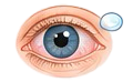 Contact-lens red eyePain, marked redness, photophobia or reduced vision in a contact-lens wearer may indicate corneal infection. Stop lens wear and arrange urgent same-day assessment.
Chemical splash: begin continuous, generous irrigation immediately, then arrange emergency assessment. Do not delay irrigation to identify the product or organise transport.
A focused first assessment
Record the information that changes urgency and helps the receiving clinician decide what to do next.
Ask about onset and risk
- Exact onset, progression, one eye or both, and whether vision is changing.
- Pain, photophobia, discharge, itch, headache, nausea, flashes, floaters, a shadow or double vision.
- Contact lenses, recent eye surgery or injection, trauma, chemical exposure and previous eye disease.
- Relevant systemic illness, skin disease, medicines, allergies and anticoagulant use.
Examine and document
- Visual acuity in each eye separately, with usual correction where available.
- Pupils, visual fields by confrontation when appropriate, eye position and movements.
- Lids and surrounding skin, conjunctiva, corneal clarity, discharge and visible foreign material.
- Compare the two eyes. Record key normal findings as well as abnormalities; do not let a normal external appearance dismiss sudden visual symptoms.
Lower the referral threshold for a new reduction in vision, significant pain, photophobia, corneal opacity, trauma, recent eye procedure or contact-lens-associated red eye.
Surface and red-eye presentations
The appearance can overlap. Pain, photophobia, corneal change and reduced vision are warning features that should prompt a different pathway.
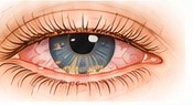
Blepharitis
Typical pattern: recurrent gritty or burning eyes, itchy or sore lid margins, and flakes or crusts around the lashes.
- Inspect the lid edges and lashes; ask about recurrence, rosacea or other skin conditions.
- Explain that symptoms can relapse. Regular gentle lid cleaning and warm compresses are common first steps.
- Reassess or refer if symptoms persist despite consistent care, the diagnosis is uncertain, or pain, photophobia or vision change occurs.
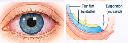
Dry-eye symptoms
Typical pattern: burning, grittiness or fluctuating blur, often in both eyes and sometimes eased by blinking.
- Ask about screen use, dry or windy environments, contact lenses and medicines that may contribute.
- Discuss environmental adjustments and appropriate lubricants; check product instructions and patient-specific factors.
- Persistent pain, photophobia, reduced vision or corneal abnormality warrants further assessment.
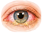
Conjunctivitis
Typical pattern: red, gritty or watery eyes; sticky discharge can occur with infection, while prominent itch often accompanies allergy.
- Check visual acuity, corneal clarity, pain, photophobia and contact-lens use before settling on a benign cause.
- Discuss hand hygiene and avoiding shared towels when infectious conjunctivitis is suspected. Treatment depends on the likely cause and local guidance.
- Urgent review for pain, photophobia, marked redness, reduced vision, corneal opacity, contact-lens use or a red sticky eye in a newborn.
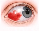
Subconjunctival haemorrhage
Typical pattern: a sharply outlined red patch on the white of the eye, usually without pain or change in vision.
- Ask about trauma, recurrent episodes, bleeding symptoms and anticoagulants; check blood pressure when appropriate.
- Confirm that vision is unchanged and there is no significant pain or photophobia.
- Investigate or refer if recurrent, unexplained, associated with trauma, or accompanied by visual symptoms. Do not stop prescribed anticoagulation without a separate clinical indication.
Red eye in a contact-lens wearer
Risk: a painful or photophobic red eye, particularly with blurred vision, needs prompt assessment for corneal disease.
- Ask about overnight wear, water exposure, lens hygiene and when symptoms began.
- Stop contact-lens wear. Do not treat a painful contact-lens red eye as routine conjunctivitis.
- Arrange urgent same-day eye assessment when pain, photophobia, reduced vision or corneal change is present.
Eyelid and tear-drainage conditions
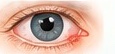
Hordeolum (stye)
Typical pattern: a focal, tender lump near the eyelid margin.
- Look for spreading redness, fever, discharge, visual change or pain in the eye itself.
- Warm compresses may help a small uncomplicated lesion. Advise the patient not to squeeze or pierce it.
- Review if it is very painful, spreading, affecting vision, recurrent or not resolving.
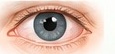
Chalazion
Typical pattern: a firm, often less-tender lump within the eyelid, related to a blocked gland.
- Check whether it is acutely inflamed and whether vision is affected.
- Warm compresses and gentle lid massage are commonly used conservative measures.
- Refer for a persistent or recurrent lump in the same place, lash loss, atypical appearance or visual effect.

Dacryocystitis
Typical pattern: tenderness, redness and swelling near the inner canthus, often with watering or discharge.
- Assess for fever, spreading inflammation, systemic illness, proptosis, restricted movements or reduced vision.
- Arrange prompt assessment through the local pathway, especially for a child, fever, systemic illness or suspected abscess.
- Systemic illness, proptosis, diplopia, reduced vision or painful/restricted movements requires emergency escalation.
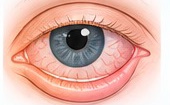
Ectropion
Typical pattern: the eyelid turns outward, which can cause watering, irritation or exposure.
- Assess surface comfort and corneal exposure; ask about persistent watering and skin irritation.
- Arrange routine eye-care review if symptoms persist or lid position affects the ocular surface.
- Pain, photophobia, corneal damage or reduced vision needs urgent ophthalmic advice.
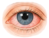
Entropion
Typical pattern: the eyelid turns inward and lashes may rub the eye, causing watering and foreign-body sensation.
- Check the cornea and visual acuity; ask about pain, photophobia and duration.
- Arrange ophthalmic assessment when lashes contact the eye or symptoms continue.
- Corneal staining or opacity, significant pain, photophobia or reduced vision requires urgent review.
Injury and sudden visual symptoms
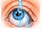
Chemical eye injury
Time-critical: pain, redness, watering and blurred vision may follow exposure to household, workplace or industrial products.
- Start continuous irrigation immediately; remove contact lenses if this can be done readily while irrigating.
- Continue emergency assessment and follow local chemical-injury protocol; bring the product details if available.
- Do not delay irrigation for history-taking, pH supplies or transport arrangements.

Corneal abrasion or foreign body
Typical pattern: pain, watering, light sensitivity and a foreign-body sensation after a scratch or particle exposure.
- Clarify mechanism and speed; check visual acuity and inspect for a penetrating injury if trained and equipped.
- High-speed injury, embedded material, reduced vision or severe pain needs urgent assessment.
- Contact-lens-associated abrasion or suspected corneal ulcer requires urgent eye-service advice.
New flashes, floaters or a curtain
Concern: sudden new floaters or flashes with a curtain, shadow or missing field can signal retinal tear or detachment.
- Ask about onset, one or both eyes, progression and any missing part of the visual field.
- Arrange urgent eye-service assessment according to the local pathway, even if the eye is painless.
- Sudden substantial vision loss requires emergency assessment.
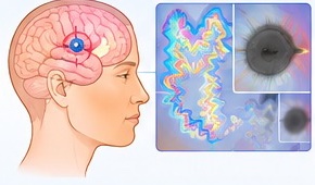
Migraine with visual aura
Possible pattern: shimmering, zigzag or expanding visual phenomena may occur with migraine, but a first or unusual episode should not be assumed to be migraine.
- Clarify whether symptoms affect one eye or the visual field, how they began and whether they fully resolved.
- New monocular vision loss, persistent deficit, sudden severe headache, weakness, speech difficulty or other neurological signs needs urgent medical assessment.
- Use the appropriate local pathway when the history is new, atypical or uncertain.
Eye drops and medicines: practical safety checks
Administration and storage
- Confirm the medicine, eye, dose schedule and duration against the prescription.
- Ask patients to wash their hands and avoid touching the bottle tip to the eye, lashes or skin.
- Check storage and discard-after-opening instructions for the individual product; allow an interval between different drops as directed.
Choose treatment deliberately
- Eye preparations include lubricants, anti-allergy medicines, antimicrobials and diagnostic agents; the choice depends on cause and local guidance.
- Check allergies, other medicines, age, pregnancy status where relevant, and ability to instil drops.
- Do not use topical anaesthetic, steroid or antibiotic drops as an empirical repeat prescription without an appropriate clinical indication and follow-up plan.
Clinical boundary: a photograph or symptom list cannot confirm a diagnosis. This guide supports triage and learning; it does not replace examination, local formularies or specialist advice.
Clinical references
The material is adapted into original wording from the supplied GCEO guide and linked to current institutional guidance. Referral timelines and pathways vary by location.
For clinician education: adapt every decision to the patient in front of you, local referral pathways and current prescribing guidance.